English
Coherent Raman Microscopy
APE & Coherent Raman Microscopy
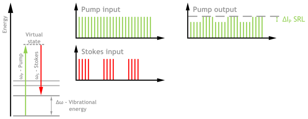
Stimulated Raman Scattering (SRS)
Stimulated Raman Scattering (SRS) microscopy for non-
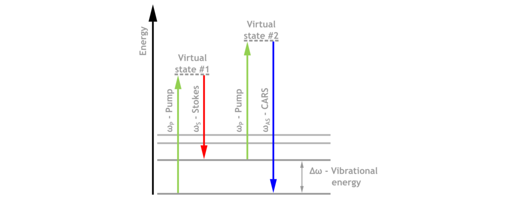
Coherent Anti-Stokes Raman Spectroscopy (CARS)
CARS Coherent anti-Stokes Raman Spectroscopy (as well as Stimulated Raman Scattering) create high imaging contrast without labeling. The technology involves two laser beams. CARS is sensitive to the vibrational modes of samples and visualizes the vibrational contrast of molecules. The samples, even living objects, remain almost unaffected.
Light Sources for CARS/SRS Microscopy
picoEmerald FT
Fast Tuning, High Sensitivity
One-Box, Turn Key and Robust
40 MHz, 660 – 2340 nm
Compatible with many Microscopes
Modular System
Customized Solutions
Flexible Design and Adaption to Special Requirements
Levante Emerald, EOM, etc.
Background Information
Among the various modalities in nonlinear optical microscopy Coherent Raman Scattering (CRS) is a very powerful one, because it enables a way to nondestructive and label-free chemical imaging.
In comparison to spontaneous Raman imaging which is using the same physical effect the CRS signal is amplified by several orders of magnitude due to coherence and stimulated effects. As a nonlinear process it offers the capability of 3D sectioning by simply shifting the focal plane within the sample.
The Raman effect describes an inelastic scattering of photons in matter, resulting in the generation of new, frequency shifted signals. This frequency shift is caused by the excitation energies of the interacting molecule. Thus it carries a signature of the molecular structure.
The resulting speed, resolution and chemical specificity makes CRS a great tool for biological imaging. Due to the fact that it does not require any labelling (sample preparation) it completes or even outperforms the common 2-photon fluorescence imaging (TPF) mode.
In the two main flavours of CRS the sample is shone with two pulsed laser beams of different wavelengths with an energy difference equal to the observed Raman transition (Pump and Stokes).
Coherent Anti-Stokes Raman Scattering (CARS)
- Four wave mixing process amplifying the Anti-Stokes wavelength
- Distorted spectral shape because of interference with non-resonant background from competing interaction channels
- Low contrast due to non-resonant background
- Signal not linear to concentration –> difficult quantitative analysis
- Easy implementation of forward- and epi-detection setup
- Imaging artefacts may appear
- Straightforward detection after spectral separation
Stimulated Raman Scattering (SRS)
- Stimulated energy transfer generating an intensity loss (SRL) and intensity gain (SRG) of the respective laser beam
- Clean Raman spectral shape
- Nearly background-free process, resulting in an intrinsic higher contrast
- Signal linear to concentration –> direct quantitative analysis
- Detection of small intensity changes requires a low-noise laser and a more elaborate detection scheme (modulation + lock-in)
- Forward detection straightforward, epi more difficult to detect